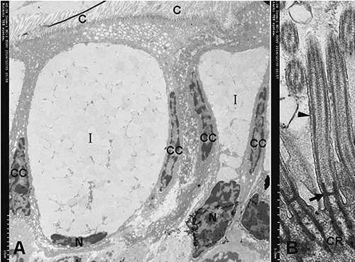
Figure 4.

A) Structure of type I mucous cells (ELV-MC), columnar cell (CC). B) Detail of the cilia (C) are inserted in narrow sags of the cell apex by two electrondense cilialy rootlets (CR); two CR are connected by an electron-dense basal plate (arrow); two CR and a basal plate form a ´H´ type frame to facilitate fixation of axoneme; the ciliary axoneme extends from the basal plate. Arrowhead, ciliary membrane.
