
Figure 2.
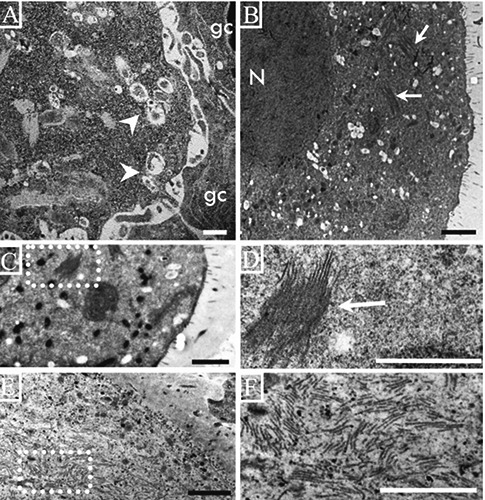
Electron micrographs of oocytes from rats of different ages. A) Deformed oocyte on a secondary follicle from a 13-day-old rat with numerous autophagic vesicles (arrow heads) but no lamellae in the cytoplasm; the granulosa cells (gc) are weakly-bound to the cytoplasm of the oocyte. B) An oocyte on an antral follicle from a 14-day-old rat; numerous vacuoles and groups of lamellae are present in the cytoplasm (arrows). C) Low magnification of the cytoplasm of an atretic oocyte on an antral follicle from a 15-day-old rat; there is a group of lamellae in the cytoplasm; the dotted square is enlarged in D. D) A group of lamellae forming a lattice structure. E) Numerous lamellae in the cytoplasm of an altered oocyte in an antral follicle from an adult rat; the lamellae are more abundant in the large, altered oocytes. F) High magnification of the dotted square in E showing depolymerized lamellae. Scale bars: 2 µm.