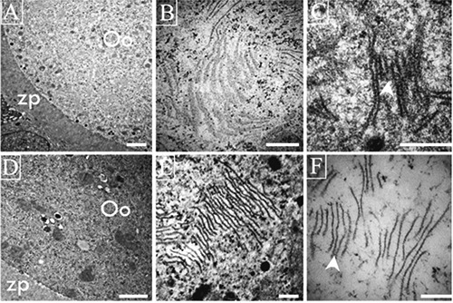
Figure 3.

Ultrastructural views of lamellae in different oocytes from antral follicles of adult rats. A-C) Lamellae in healthy oocytes form lattices; in C one can observe the strong relation among the several lamellae units that form lattices (arrow head). D-F) Altered oocyte shows depolymerized lamellae present as fibrous units visible under high magnification (F) of zones of the same oocytes illustrated in D (arrow head). Scale bars: A,D) 2 µm; remaining panels: 200 nm.
