
Figure 5.
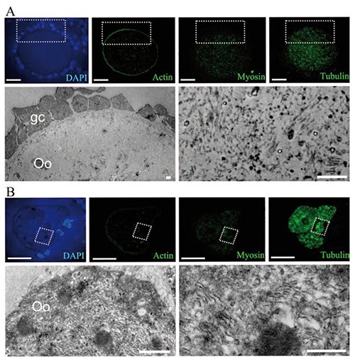
Optical immunodetection of the proteins actin, myosin and tubulin in a normal oocyte (A), and an altered oocyte (B), with ultrastructural evaluation on antral follicles. A) The normal oocyte displays a peripheral pattern of actin distribution, while myosin and tubulin are distributed in the cytoplasmic space; the electron microscopy image shows the square area illustrated in the semi-thin sections, evidencing the small amount of lamellae that corresponds to the low quantity of immunolabeling. B) A highly-altered, segmented oocyte with a high amount of tubulin; the ultrastructure of the squared zone is shown in the lower panel, evidencing a high amount of depolymerized lamellae. Scale bars: optical images, 20 m; electron microscopy images, 2 µm.