Figure 8.

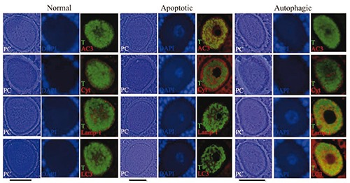
Simultaneous light immunolocalization of tubulin with apoptotic and autophagic proteins, in continuous sections of paraffin-embedded samples on oocytes from antral follicles. Phase contrast illumination (PC) evidences the different morphology corresponding to normal and altered oocytes (apoptotic and autophagic). In the normal oocyte the green label shows tubulin (T) distributed homogenously in the cytoplasmic space that coincides with a basal label to cytochrome-C (Cyt), Lamp-1 and LC3 proteins as well as the negative label to active caspase-3 (AC3). In the apoptotic oocyte the aggregated tubulin distribution coincides with an increased label to cytochrome-C and the positive reaction to the active caspase-3. In the autophagic oocyte the tubulin is distributed homogenously in the cytoplasmic space and does not form aggregates. This oocyte has an increased level of Lamp-1 and LC3 proteins. DAPI is evidencing the chromatin distribution in the nuclear space. Scale bars: 30 µm.
