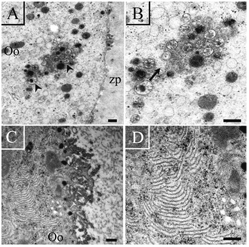
Figure 9.

Ultrastructural visualization of altered oocytes from antral follicles. A) Autophagic vesicles (arrow) are present in the cytoplasm of the oocyte (Oo); some cytoplasmic prolongations of the granulosa cells are observed (arrow heads). B) High magnification of the same oocyte observed in A; the autophagic vacuoles are indicated by an arrow; the oocyte has no lamellae. C) Region of a fragmented oocyte with a high quantity of lamellae. D) High magnification of the same oocyte shown in C; the presence of depolymerized lamellae is observed in its cytoplasm. Scale bars: left panel, 2 µm; right panel, 500 nm.
