
Figure 10.
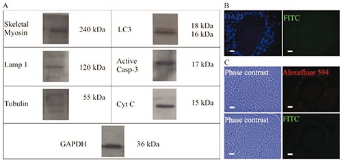
Western blot analysis and negative controls for the different light immunolocalizations performed. A) Analyses of total myosin, Lamp 1, tubulin, (LC3-I and LC3-II), active caspase-3, and cytochrome-C from isolated oocytes; the presence of the different proteins evaluated was evidenced in the Western Blot assays using total proteins from isolated oocytes. B) Negative control to the immunodetection realized in semi-thin sections. C) Negative control to the immunodetection realized in sectioned paraffin-embedded samples. Scale bars: 10 µm.