
Figure 3.
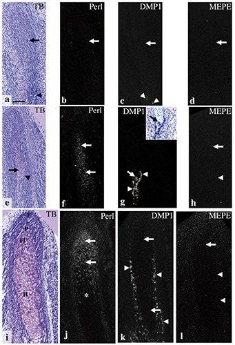
Condylar anlage/cartilage in coronal plane at E14.0 (a-d), E15.0 (e-h), and E16.0 (i-l). Toluidine blue staining (a,e,i), and in situ hybridization for perlecan (b,f,j), DMP1 (c,g,k), and MEPE (d,h,l). Inset in (g) shows a bright field image of identical section stained with toluidine blue. a) Although the ossifying mandible was recognizable (arrowhead), metachromasia was not observed in the condylar anlage (arrow). b-d) Perlecan, DMP1, and MEPE mRNA expression were not detected in the anlage (arrows in b-d), but DMP1 mRNA was slightly expressed in cells in the osteoid-like tissue (arrowheads in c). e) A metachromatically stained matrix was first detected in the anlage (arrow); note that newly formed chondrocytes showed a considerable hypertrophy, and the bone collar had formed around the cartilage (arrowhead). f) Perlecan mRNA expression was detected in newly formed chondrocytes (arrows). g) DMP1 mRNA was expressed in the osteogenic cells of the bone collar (arrowheads) as well as in a few chondrocytes (arrows). h) MEPE mRNA expression was barely detected both in the cartilage (arrow) and the bone collar (arrowhead). i) Cell zones in the condylar cartilage had become distinct: fibrous cell (articular cell) zone (F), polymorphic cell zone (P), flattened cell zone (Fl), and hypertrophic cell zone (H). j) Perlecan mRNA was expressed in chondrocytes from flattened cell zone to upper hypertrophic cell zone (arrows), but reduced in the lower hypertrophic cell zone (*). k) DMP1 mRNA was expressed in osteogenic cells of the bone collar (arrowheads), but not in chondrocytes (arrows). l) MEPE mRNA expression was not detected in the bone collar (arrowheads) or in the cartilage (arrow). Scale bars: 100 µm.