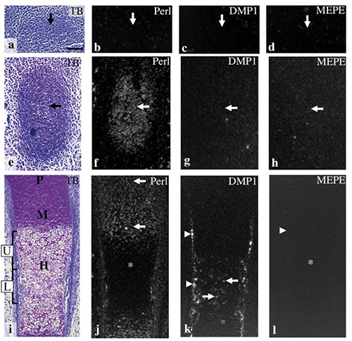
Figure 4.

The anlage of the future tibia at E12.0 (a-d), and tibial cartilage at E13.0 (e-h), E14.0 (i-l). Toluidine blue staining (a,e,i) and in situ hybridization for perlecan (b,f,j), DMP1 (c,g,k), and MEPE (d,h,l). a) The anlage of the future tibia consisted of a mesenchymal cell condensation (arrow). b-d) Perlecan, DMP1, and MEPE mRNA were not expressed in the condensation (arrows). e) A metachromatically stained matrix was first detected in the anlage of the future tibia (arrow). f) Perlecan mRNA was expressed in chondrocytes throughout the cartilage (arrow). g,h) DMP1 and MEPE mRNA were not detected in tibial cartilage (arrows). i) Proliferative (P), maturation (M), and hypertrophic cell zone (H) had become clearly identifiable in the cartilage, and hypertrophic cell zone could be divided into two zones: the upper hypertrophic cell zone (U) and the lower hypertrophic cell zone (L). j) Perlecan mRNA was expressed in chondrocytes throughout the entire proliferative and maturation cell zone (arrows), but expression was weaker in the hypertrophic cell zone (*). k) DMP1 mRNA was expressed in the osteogenic cells of the bone collar (arrowheads) and in chondrocytes of the lower hypertrophic cell zone (arrows). l) MEPE mRNA expression was not detected both in the bone collar (arrowhead) or in the cartilage (*). Scale bars: 100 µm.
