Figure 5.

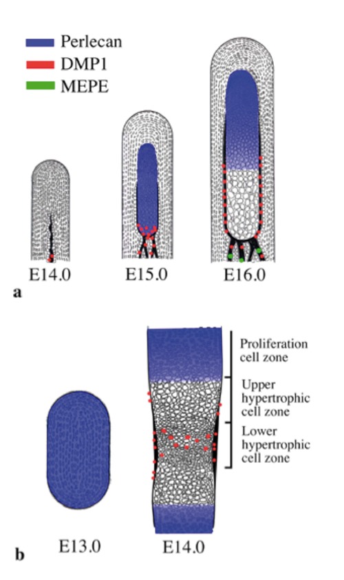
Schematic representations of the expression pattern of perlecan, DMP1, and MEPE based on the present findings. a) Condylar anlage/cartilage at E14.0 to E16.0. b) Tibial cartilage at E13.0 and E14.0.

Schematic representations of the expression pattern of perlecan, DMP1, and MEPE based on the present findings. a) Condylar anlage/cartilage at E14.0 to E16.0. b) Tibial cartilage at E13.0 and E14.0.