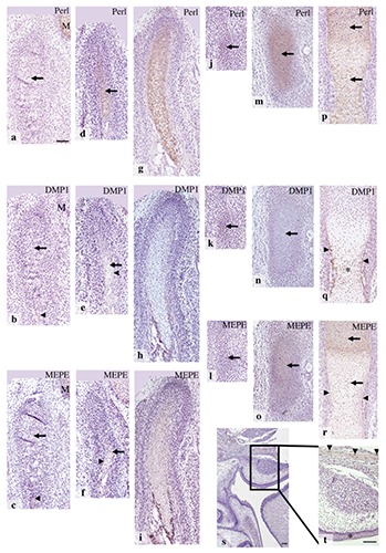
Figure 6.

Condylar anlage/cartilage at E14.0 (a-c), E15.0 (d-f), E16.0 (g-i), and maxillary region at E16.0 (s,t); t) is the higher magnification of rectangular area of (s). The anlage of future tibia at E12.0 (j-l), and tibial cartilage at E13.0 (m-o), and E14.0 (p-r). Immunohistochemistry for perlecan (a,d,g,j,m,p), DMP1 (b,e,h,k,n,q), and MEPE (c,f,i,l,o,r,s,t). a) Perlecan immunoreactivity was evident in Meckel’s cartilage (M), but not in the anlage (arrow). b) DMP1 immunoreactivity was weak but definite in the osteoid-like tissue (arrowhead), but not in the condylar anlage (arrow) or Meckel’s cartilage (M). c) MEPE immunoreactivity was not detected in the condylar anlage (arrow), but it was evident in the osteoid-like tissue (arrowhead) and in Meckel’s cartilage (M). d) Perlecan immunoreactivity was clearly detected in newly formed cartilage matrix (arrow). e) DMP1 immunoreactivity was evident in the bone collar (arrowhead), but not in the cartilage matrix (arrow). f) MEPE immunoreactivity was evident in the bone collar (arrowhead) and in the cartilage matrix (arrow). g-i) Immunostaining patterns were similar to those of E15.0. j-l) Perlecan, DMP1, and MEPE immunoreactivity were not detected in the mesenchymal cell condensation (arrows). m-o) Perlecan and MEPE immunoreactivity was evident in the matrix throughout the cartilage matrix (arrows in m and o), but DMP1 immunoreactivity was not detected (arrow in n). p) Perlecan immunoreactitivity was detected throughout the cartilage matrix (arrows). q) DMP1 immunoreactivity was detected in the bone collar (arrowheads) and in the lower hypertrophic cell zone (*). r-t) MEPE immunoreactivity was detected in the bone collar (arrowheads in r), throughout the cartilage (arrows in r), and in some bone lacunae (arrowheads in t), but not in the oral epithelium (asterisk in t). Scale bars; a-r) 100 µm; s,t) 80 µm.
