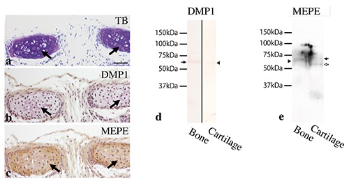
Figure 7.

Tracheal cartilage at 3 weeks old (a-c) and Western immunoblotting for DMP1 and MEPE in bone- or cartilage- derived samples (d,e). a) Tracheal cartilage matrix exhibited metachromasia by toluidine blue staining (arrows). (b) DMP1 immunoreactivity was not detected in the cartilage matrix (arrows). c) MEPE immunoreactivity was clearly recognized in the cartilage matrix (arrows). d) DMP1 antibody clearly recognized a strong protein band of 65 kDa in bone-derived samples (arrow), but a weaker band in cartilage-derived samples (arrowhead). e) MEPE antibody recognized a protein band of 62 kDa in bone-derived samples (arrowhead) and two bands, one of 67 kDa (black arrow) and another of 59 kDa (white arrow) in cartilage-derived samples. Scale bars: 50 µm.
