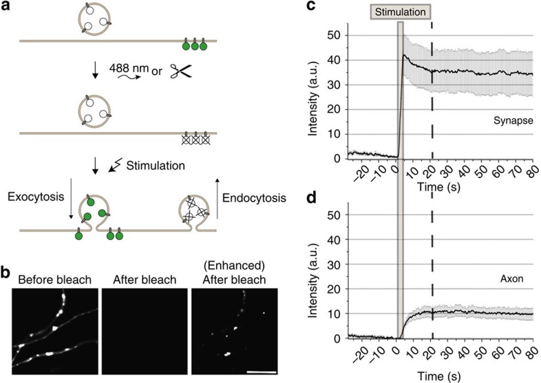
Figure 1. Newly exocytosed Syb2 is largely retained within presynaptic boutons.
(a) Schematic of experimental workflow to monitor newly exocytosed pHluorin-tagged SV proteins. (b) Syb2-pHluorin fluorescence (F) before and after selective photobleaching. Right, enhanced contrast shows small residual F post-bleaching. Scale bar, 5 μm. (c,d) Syb2-pHluorin F intensity changes from time-lapse images (400 ms frame−1) of stimulated (40 APs, 20 Hz) surface-eclipsed neurons. (c) Presynaptic boutons, (d) axonal areas (mean±s.e.m.; N=10 neurons).

