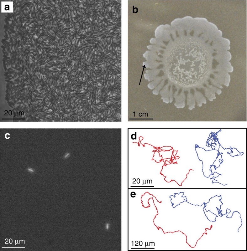
Figure 1. Tracking individual bacteria within a dense swarm.
(a–b) Phase contrast imaging of a wild type B. subtilis swarming colony: at high (a) and low (b) magnifications (region of interest is marked with an arrow in (b)). (c) Fluorescent microscopy showing the fluorescently labelled bacteria only, at high magnification. (d–e) Example trajectories of individual bacteria inside the swarm at high (d) and low (e) magnifications. Left/Red: B. subtilis and Blue/Right: S. marcescens.

