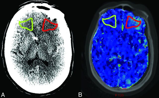
Fig 1.
ROI placement and coregistration in a representative patient. A, NCCT of a representative patient with SAH demonstrates ROI placement in the region of a new left frontal infarction, which was not present on admission (red) and the contralateral control ROI (green). B, Coregistration of the NCCT from A with the preinfarction CTP yields matched ROI placement on the PS map and the CBF and MTT maps (not shown).

