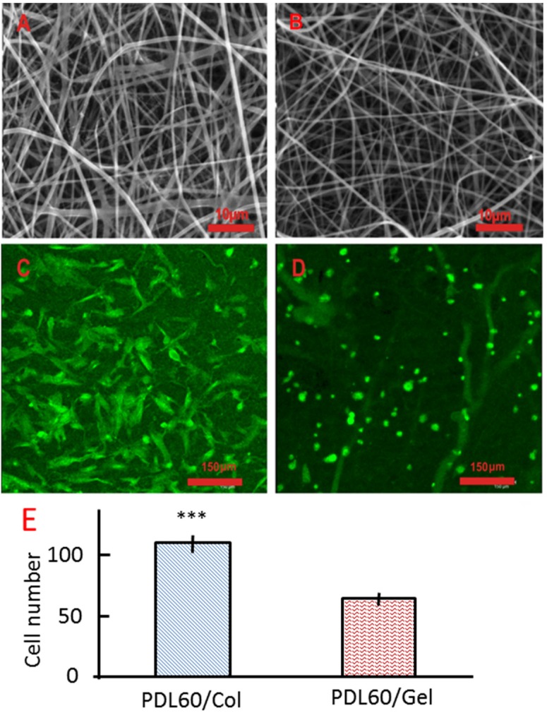
Figure 6.
HBMSCs attachment on the PDL60/Col and PDL60/Gel electrospun scaffolds. (A,B) are SEM images of PDL60/Col scaffolds and PDL60/Gel scaffolds, respectively. (C,D) are maximum projection fluorescence images of PDL60/Col and PDL60/Gel cell-scaffold constructs, respectively. Images were collected using confocal microscopy with 20× water immersion objective. (E) Cell numbers were counted based on the confocal images and given as mean ± SD (n =3), two-tail, unpaired t-test was performed, ***p < 0.001.

