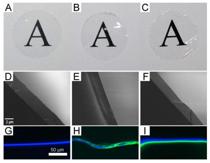
Figure 3.
Physical appearance of membranes prepared from either fibroin alone (A, D, and G), tropoelastin-fibroin blend (10:90 ratio) (B, E, and H), and layered solutions of fibroin and tropoelastin (C, F, and I); (A–C): gross appearance of each membrane when placed over printed text (16-mm diameter discs); (D–F): internal structures revealed by scanning electron microscopy following freeze-fracture; and (G–I) visualization of tropoelastin (green) by immunolabelling and confocal fluorescence microscopy (the presence of fibroin revealed as blue autofluorescence).

