Figure 3.

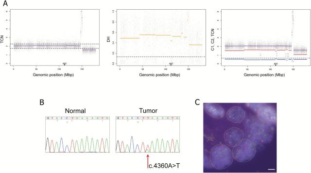
UPD of chromosome 5 accompanied by a homozygous nonsense mutation of sample 33. (A) Tumor-matched normal PSCBS profiling of SNP array data showing total copy number (panel 1), decrease of heterozygosity (panel 2) and allele-specific copy number (panel 3). Dotted horizontal lines represent specific thresholds for each parameter. As illustrated, APC is located in a region with cnLOH. (B) Sanger sequencing of the gene APC showing the existence of a nonsense mutation (c.4360A>T) in the tumor. The minority allele could represent intratumoral heterogeneity and/or infiltration of normal cells. (C) Microscopic assessment of FISH signals using fluorescent probes covering the gene APC (red) and a control probe at 5q31.2 (green). Single isolated nuclei displaying two copies of APC and the control probe confirmed that most tumor population showed a disomic chromosome 5q within this sample. Scale bar = 5 µm.
