
Figure 14. 3D image registration with the presented validation model.
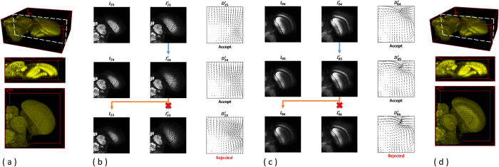
Using the serial-section laser scanning microscope images of the drosophila brain data as example, (a) 3D reconstruction results with a side view of the raw input data are displayed; (b) an example of the backward registration from the image layer I25 to the image layer I23 is presented with associated deformation fields and validation processes. As the validation model accepts the deformation fields,  , the outputs of the registration will be
  . On the other hand,
  is rejected by the validation model, and the registration output will be I23. (c) Similarly, an example of the forward registration from Layer I84 to the image layer I86 is shown with associated deformation fields and validation processes. (d) 3D reconstruction results with a side view of the registered outputs are presented.