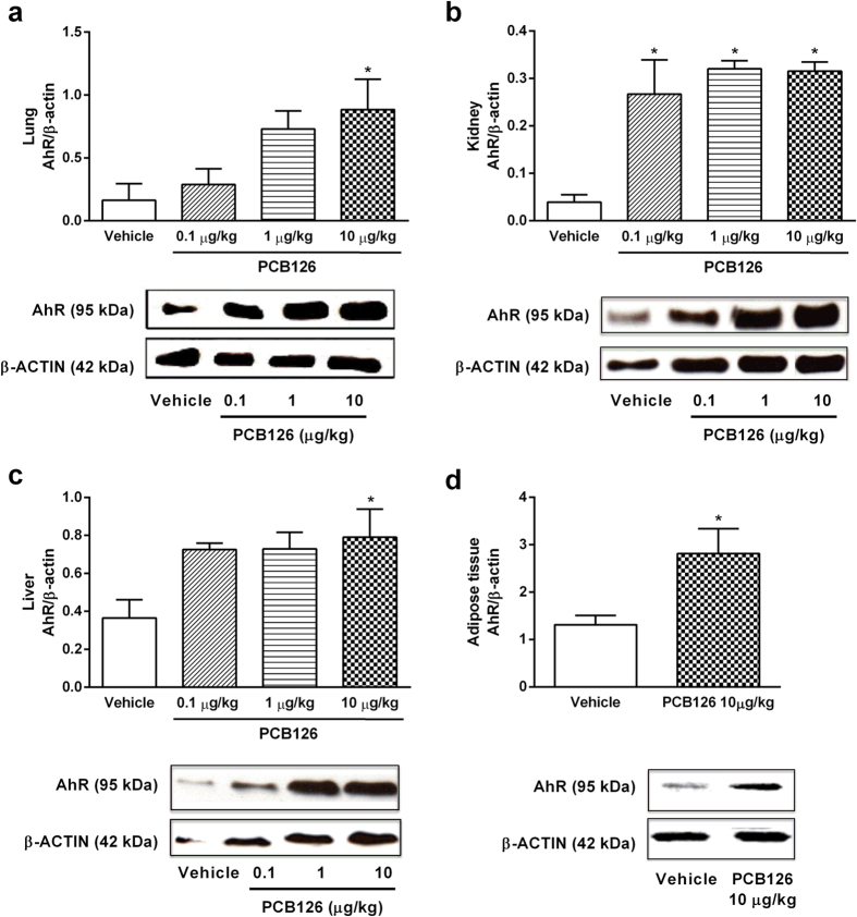
Figure 2. In vivo PCB126 exposure up-regulates AhR expression in target tissues.
AhR protein expression in the lung (a), kidney (b) and liver (c) tissues collected from male Wistar rats exposed to vehicle or PCB126 (0.1, 1, or 10 μg/kg once a day during 15 days). AhR protein expression in the adipose tissue (d) collected from male Wistar rats exposed to vehicle or PCB126 (10 μg/kg once a day during 15 days). Five hours after the last exposures, samples were collected and AhR levels were quantified by Western blot. Data (n = 4 for each group) were analysed by one-way ANOVA followed by Tukey’s test (a–c) or Student’s t-test (d). *P < 0.05 vs. vehicle.

