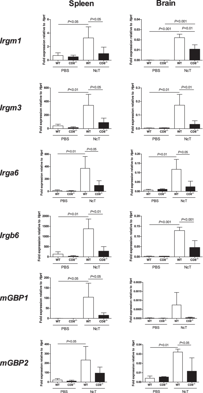
Figure 6. Lack of CD8+ T cells decreases IRG mRNA expression in infected mice.
Relative levels of Irgm1, Irgm3, Irga6, Irgb6, mGBP1 and mGBP2 mRNA, normalized to hypoxanthine guanine phosphoribosyl transferase (Hprt) mRNA, detected by real-time PCR in the spleen and brain of WT and CD8a−/− mice, as indicated, 7 days after i.p. injection of 1 × 10⁷ N. caninum tachyzoites (NcT; n = 4) or PBS (PBS; n = 3). Bars represent mean values of the respective group plus one SD. Statistical significance between infected mice and controls is indicated above bars (one-way ANOVA and Tukey’s post-hoc test).

