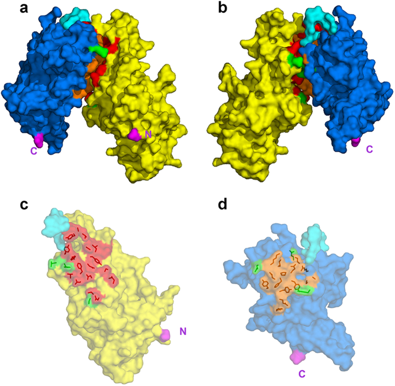
Figure 2. Interface between the DBL3X and DBL4ε domains.
The domains are shown in surface representation with DBL3X in yellow, DBL4ε in blue and the linker region in cyan. The invariant contacting residues (see Table S1) are in red for DBL3X and orange for DBL4ε; polymorphic contacting residues are green for both domains. The N- and C-termini of the double domain are in magenta and are labelled N and C. (a,b) The DBL3X-DBL4ε double domain viewed edge on from the interdomain interface. The two views are rotated by 180° with respect to each other about the vertical (c) Semi-transparent surface representation of the DBL3X domain viewed from above the interface with the side chains of the contacting residues shown in red. (d) Semi-transparent surface representation of the DBL4ε domain viewed from above the interface with the side chains of the contacting residues shown in orange.

