
Fig. 2.
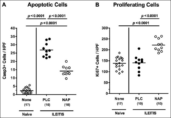
Apoptotic and proliferating cells in ileal samples of NAP-treated mice following acute ileitis induction. In order to induce acute ileitis, C57BL/6j wildtype mice were perorally infected with 100 cysts of T. gondii ME49 strain on day 0. Mice were treated either with synthetic NAP (open circles) or placebo (PLC; filled circles). Naive, uninfected, and untreated (None; open diamonds) mice served as negative controls. The average numbers of apoptotic (positive for caspase-3, Casp3, panel A) and proliferating cells (positive for Ki67, panel B) from at least six high power fields (HPF, 400× magnification) per animal were determined microscopically in immunohistochemically stained ileal paraffin sections at day 7 p.i. Numbers of analyzed animals are given in parenthesis. Medians (black bars) and level of significance (p-value) determined by Mann–Whitney U test are indicated. Data were pooled from three independent experiments