
Fig. 3.
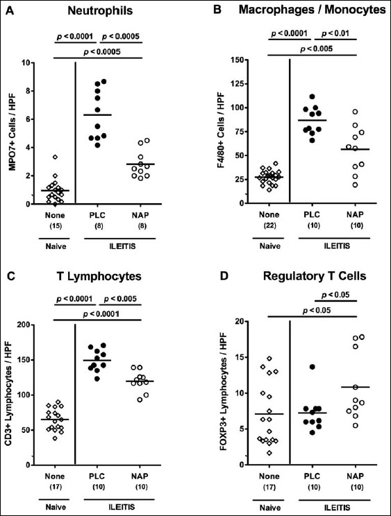
Ileal immune cell responses in NAP-treated mice following acute ileitis induction. In order to induce acute ileitis, C57BL/6j wildtype mice were perorally infected with 100 cysts of T. gondii ME49 strain on day 0. Mice were treated either with synthetic NAP (open circles) or placebo (PLC; filled circles). Naive, uninfected, and untreated (None; open diamonds) mice served as negative controls. The average number of cells positive for (A) MPO7 (neutrophils), (B) F4/80 (macrophages and monocytes), (C) CD3 (T lymphocytes), and (D) FOXP3 (regulatory T cells) from at least six high power fields (HPF, 400× magnification) per animal was determined microscopically in immunohistochemically stained ileal paraffin sections derived from mice at day 7 p.i. Numbers of analyzed animals are given in parenthesis. Medians (black bars) and level of significance (p-value) determined by Mann–Whitney U test are indicated. Data were pooled from three independent experiments