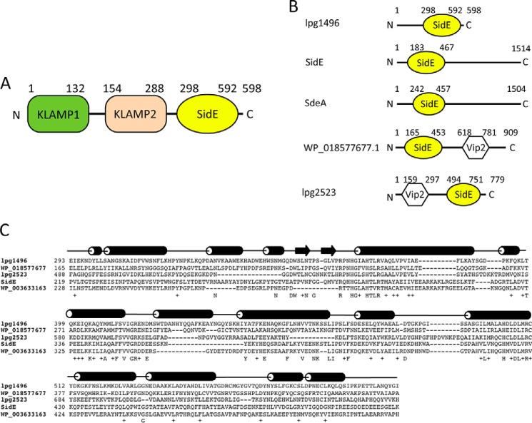
FIGURE 1.
SidE sequence and structure. A, domain architecture of lpg1496 (KLAMP1 in green, KLAMP2 in wheat, SidE in yellow). B, the occurrence of the SidE domain in Legionella proteins. C, sequence alignment of the SidE domain of lpg1496 with other bacterial proteins. The secondary structure of the lpg1496 SidE domain is overlaid on top (cylinder = α-helix; arrow = β-sheet).

