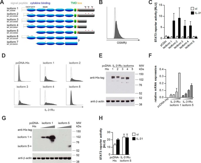
FIGURE 1.
STAT3 activation by human IL-31Rα isoforms. A, schematic representation of the domain architecture of the seven human IL-31Rα isoforms. The variable portion of the intracellular domain is shown in gray. Gray marks indicate cysteine residues in the cytokine binding domain. Tyrosine residues implicated in STAT3 recruitment are highlighted in red. TMD, transmembrane domain; box, JAK-binding box. B, flow cytometric analysis of human OSMRβ expression in HeLa cells. The white histogram indicates the isotype control. C, luciferase reporter gene assay. HeLa cells were transfected with a STAT3 luciferase reporter, a Renilla-luciferase expression plasmid, and expression plasmids encoding human IL-31Rα isoforms. As a control an equal amount of the empty vector (pCDH) was co-transfected. One day post-transfection, cells were treated with rhIL-31 (30 ng/ml). Luciferase activity was assessed after 24 h of cytokine stimulation. Data represent mean values of five independent experiments carried out in duplicate, normalized to the corresponding Renilla luciferase signals. White bars, uninduced (ui); black bars, IL-31. Error bars indicate standard deviations. RLU, relative light units. D, flow cytometric analysis of different IL-31Rα isoforms transfected into HeLa cells. White histograms display the isotype control. E, Western blotting. HeLa cells were transfected with expression plasmids encoding His-tagged human IL-31Rα isoforms or empty pcDNA-His expression vector. IL-31Rα protein expression was detected with His tag-specific antibodies. Expression of β-actin was monitored to control for equal loading. F and G, mRNA and protein expression of IL-31Rα isoforms 1 and 5 in HeLa cells. Cells were transiently transfected with increasing amounts of plasmids encoding human IL-31Rα isoforms 1 and 5 (gray triangles). As a control, the empty vector (pcDNA-His) was transfected. mRNA expression of IL-31Rα isoforms 1 and 5 was analyzed by qRT-PCR (F). Protein expression of IL-31Rα isoform 1 and isoform 5 is shown by Western blotting using an anti-His tag antibody (G). β-Actin expression was monitored to control for equal loading. H, STAT3 reporter gene assay. HeLa cells were transiently transfected with a STAT3 luciferase reporter and IL-31Rα isoform 5 at the concentration leading to detectable protein expression as shown in G. 24 h post-transfection, cells were stimulated with rhIL-31 (30 ng/ml). Luciferase activity was assessed 24 h after cytokine stimulation. Error bars indicate standard deviations. ALU, arbitrary light units.

