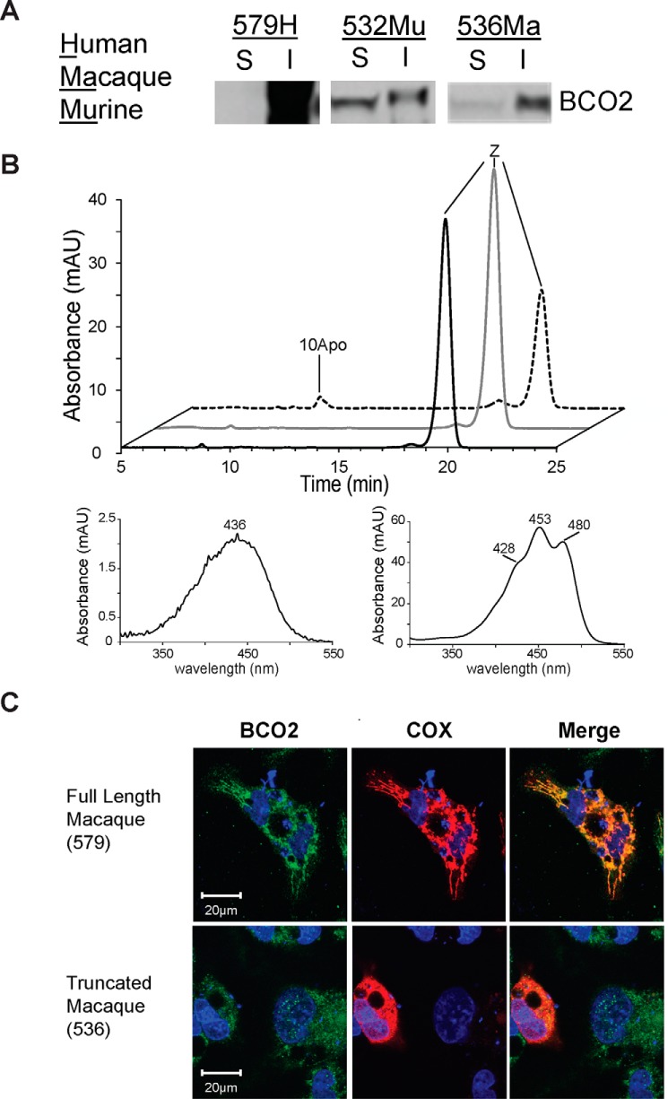
FIGURE 3.
Recombinant expression, enzyme activity, and cellular localization of BCO2s. A, Western blot of soluble (S) and insoluble (I) centrifugal fractions from lysed bacteria expressing recombinant (from left to right) human, murine, and macaque BCO2. B, insoluble fraction of HuBCO2 expressed in E. coli is inactive. HPLC of lipid extracts of in vitro enzyme activity assays of soluble MuBCO2 (dashed trace), insoluble MuBCO2 (black trace), and insoluble HuBCO2 (gray trace) on zeaxanthin was carried out. UV spectra of 10Apo and zeaxanthin are shown. C, confocal images of HepG2 cells transfected with full-length (top panel) and truncated (bottom panel) macaque BCO2-encoding plasmids. Immunostaining was performed with anti-V5 antibody for BCO2 (red) and anti-cytochrome c oxidase subunit IV (COX) antibody (green). Nuclei were stained with DAPI (blue). Only the full-length MaBCO2 shows co-localization with cytochrome c oxidase subunit IV in merged images. mAU, milli-absorbance units.

