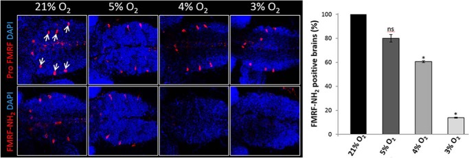
FIGURE 8.

Immunofluorescence of Drosophila melanogaster 3rd instar larval brains with antibodies to the FMRF pro-peptide (Pro FMRF) or to the amidated product, FMRF-NH2, merged with DAPI images. Drosophila 1st instar larvae were developed under different oxygen concentrations and dissected at late 3rd instar for immunostaining. Arrows indicate Tv neurons, which are positive for FMRF (n > 30 brains in three independent experiments, Student's t-test. *, p < 0.001; ns: non-significant; bars represent S.E.). DAPI labels cell nuclei (blue).
