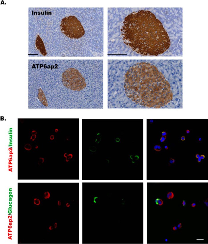
FIGURE 4.
ATP6ap2 expression in the pancreas and islets. A, immunohistochemistry showing ATP6ap2 localization in the mouse pancreatic sections. The right panels show the enlarged images of the left panels. Bar, 100 μm. B, immunofluorescence showing ATP6ap2 localization in dispersed human islets. Bar, 20 μm. Representative images are from three independent experiments.

