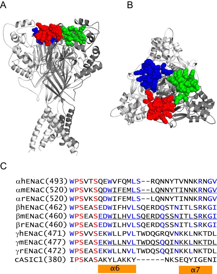
FIGURE 1.
Knuckle domain location and sequence alignments. A, chicken ASIC1 structural model showing knuckle domain locations in the channel complex. The trimeric cASIC1 model was built from structural coordinates of the minimal functional cASIC1 (PDB entry 4NYK superseding 3HGC (48)). The three cASIC1 subunits are rendered as solid ribbons in dark, intermediate, and light gray using PyMOL (49). The knuckle domain residues of three subunits are rendered as space-filled spheres in red, blue, or green. B, top view of the same model in A. C, sequence alignments of human, mouse, and rat ENaC subunits and cASIC1. Alignments were done using Vector NTI 11 (Invitrogen). Identical residues are in red, and conserved residues are in blue. Mouse ENaC residues that were deleted in this study are underlined. Chicken ASIC1 residues within the α6 and α7 helices are indicated by two orange bars.

