
FIGURE 3.
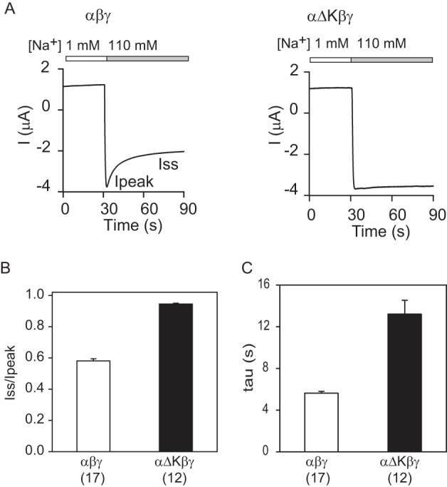
α subunit knuckle domain deletion results in a loss of Na+ self-inhibition. A, representative traces for Na+ self-inhibition responses in oocytes expressing αβγ and αΔKβγ. Oocytes were clamped at −60 or −100 mV, and whole-cell currents were continuously recorded while bath [Na+] was rapidly increased from 1 mm (open bar) to 110 mm (gray bar). Inward currents are shown as negative values by convention. The current decline following the increase in bath [Na+] from a peak current (Ipeak) to a steady-state current (Iss) represents Na+ self-inhibition. B, Na+ self-inhibition magnitudes (Iss/Ipeak) of αβγ and αΔKβγ. The Iss/Ipeak values were obtained as described under “Experimental Procedures.” Data were collected in three batches of oocytes with the total oocyte numbers shown in parentheses. C, Na+ self-inhibition time constants of αβγ and αΔKβγ. The Iss/Ipeak ratio and τ of αΔKβγ were significantly greater than that of WT (black columns, p < 0.001). Data were collected in three batches of oocytes. Total oocyte numbers are shown in parentheses below. Error bars, S.E.