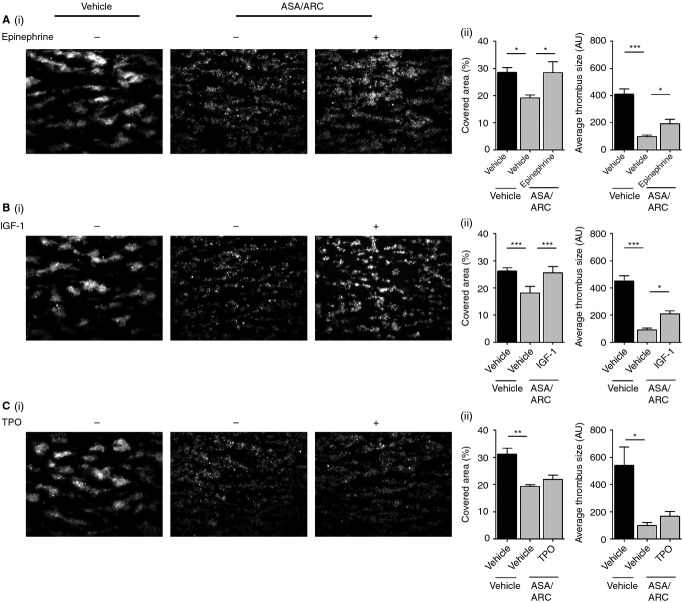
Figure 6.
Ex vivo thrombus formation on collagen is reduced by dual antiplatelet therapy, a process that is rescued by insulin-like growth factor-1 (IGF-1) and epinephrine. Fluorescently labeled (dihexyloxacarbocyanine iodide [DiOC6]) whole blood anticoagulated with 2 U mL−1 heparin and 40 μm d-phenylalanylprolyl-arginyl chloromethyl ketone was pretreated with vehicle control or 1 μm AR-C66096 (ARC) and 30 μm acetylsalicylic acid (ASA) in combination for 10 min. Blood samples were subsequently preincubated with vehicle control or primer, i.e. 100 nm epinephrine (A), 100 nm IGF-1 (B), and 50 ng mL−1 thrombopoietin (TPO) (C), as indicated for 5 min. Samples were then perfused at an arterial shear rate of 1000 s−1 over fibrillar collagen (50 μg mL−1) for 5 min. Samples were washed with HEPES–Tyrode buffer for 2 min to remove non-adherent cells. Representative fluorescent images are shown, along with quantitative analysis of surface area covered (%) with thrombi and the average thrombus size (AU) Data represent the average results taken from ≥ 15 random microscopic fields per experiment (n = 4–5). Statistical analysis: one-way anova was used in conjunction with a Bonferroni post hoc test; *P < 0.05, **P < 0.01, ***P < 0.001.

