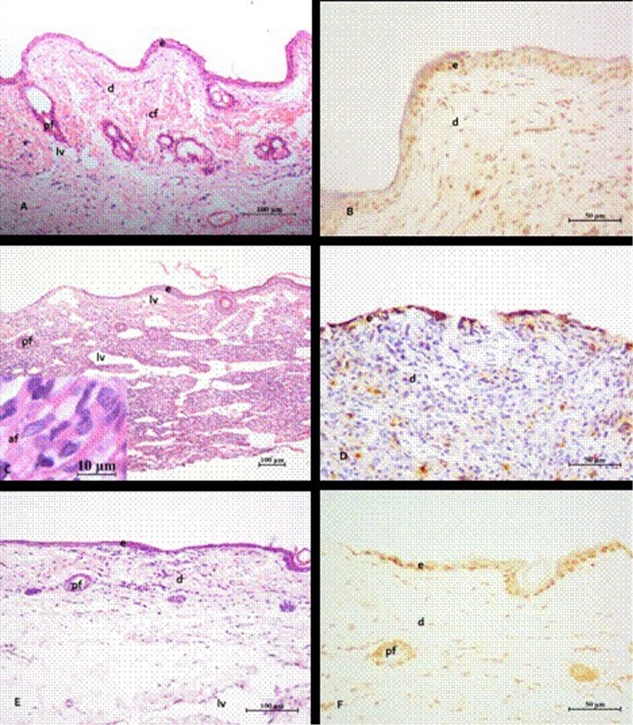
Figure 4 .

Photomicrograph of the footpad lesions of hamsters infected with Leishmania braziliensis. Lesions treated with MB/LED-PDT with methylene blue (MB) administered topically (Tp) (A and B). The control animals (C and D) were untreated or were treated with amphotericin B (AmB) (E and F). After MB/LED-PDT (A), intact skin with hair follicles (pf) in regeneration, absence of inflammation and few dilated lymphatic vessels (lv) are noted. The dermis (d) showed thick bundles of collagen fibers (cf). In infected untreated animals (C), intense inflammatory infiltrate intermingled with dilated lymphatic vessels are noted. These lesions showed partial loss of epidermis (e) and frequent occurrence of amastigote forms (af) (detail in C) in the dermis region. After treatment with AmB (E), regenerated epidermis and dermis in the repair process, with beams of smaller and thinner collagen fibers are noted. In (B, D and F) the sections stained with TUNEL showed that higher frequency of labeled cells, particularly fibroblasts and keratinocytes occurred in lesions treated with MB/LED-PDT, followed by those treated with AmB and those not treated. Stain: hematoxylin and eosin (HE) (A, C and E); TUNEL (B, D and F).
