Figure 6 .

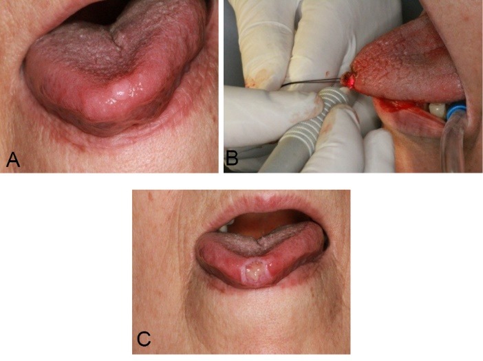
(A) A nodular exophytic lesion with smooth surface on anterior part of dorsal surface of tongue. (B) Lesion was excised with diode laser. (C) Seven days after procedure we saw an ulcer with fibrino-leucocytaire-pseudomembrane on surface.

(A) A nodular exophytic lesion with smooth surface on anterior part of dorsal surface of tongue. (B) Lesion was excised with diode laser. (C) Seven days after procedure we saw an ulcer with fibrino-leucocytaire-pseudomembrane on surface.