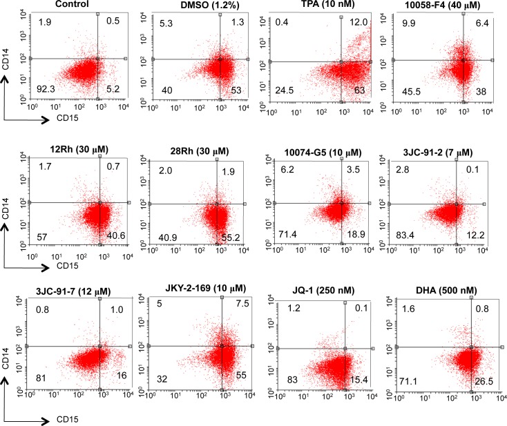
Figure 2. Myc inhibitors promote myeloid differentiation of HL60 cells.
HL60 cells in log-phase growth (ca. 105 cells/ml) were incubated with the indicated concentrations of Myc inhibitors for 4-5 days at which point they were stained with mAbs directed against cell surface CD14 and CD15. Separate cultures were incubated with DMSO or 12-O-tetradecanoylphorbol-13-acetate (TPA), as controls for “pure” myeloid and monocyte/macrophage differentiation, respectively. Cell surface fluorescence was evaluated by two-color flow cytometry.

