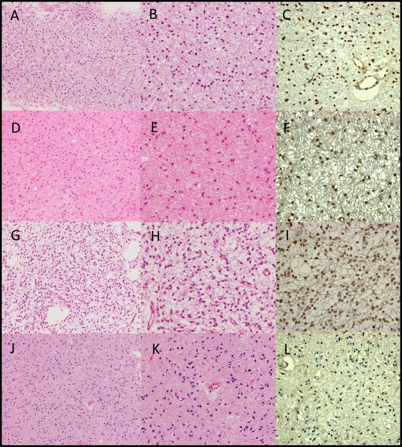
Figure 4. Hematoxylin and eosin staining (a, b, d, e, g, h, j, k) and ATRX staining (c, f, i, l) of examples of “astrocytic” gliomas with total 1p19q loss.
a, b, c. Table S1, case 39. Institutional diagnosis: diffuse astrocytoma. Single-pathologist diagnosis: diffuse astrocytoma. d, e, f. Table S1, case 44. Institutional diagnosis: diffuse astrocytoma. Single-pathologist diagnosis: diffuse astrocytoma. g, h, i. Table S1, case 52. Institutional diagnosis: anaplastic astrocytoma. Single-pathologist diagnosis: diffuse astrocytoma. j, k, l. Table S1, case 53. Institutional diagnosis: anaplastic astrocytoma. Single-pathologist diagnosis: diffuse astrocytoma. a, d, g, j: original magnification ×100. b, c, e, f, h, i, k, l: original magnification ×200.

