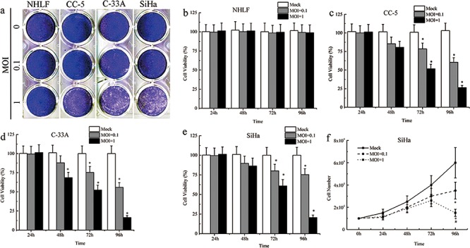
Figure 2. Cytopathic effects and cell death induced by MV-Edm.

a. Serial analysis to determine the cytopathic effects (CPEs) of MV-Edm was performed on the human CC cell lines SiHa and C-33A, primary cancer cells CC-5 and normal cell line NHLF. Ninety-six hours after infection at MOIs of 0.1 and 1, the cells were stained with crystal violet representing viable attached cells. b–e. The time course of cell viability of the human CC cell lines, primary cancer cells and normal cell line after infection with MV-Edm at MOIs of 0.1 and 1 was analyzed by the MTS assay. f. Live Cell counts assay was performed to evaluate the number of cells by Trypan Blue Staining. All data were presented as means ± SD. *means P < 0.05 when compared to MOCK group.
