
Figure 4. DDR1 expression affects IGF-I mediated biological effects in human cancer cells.
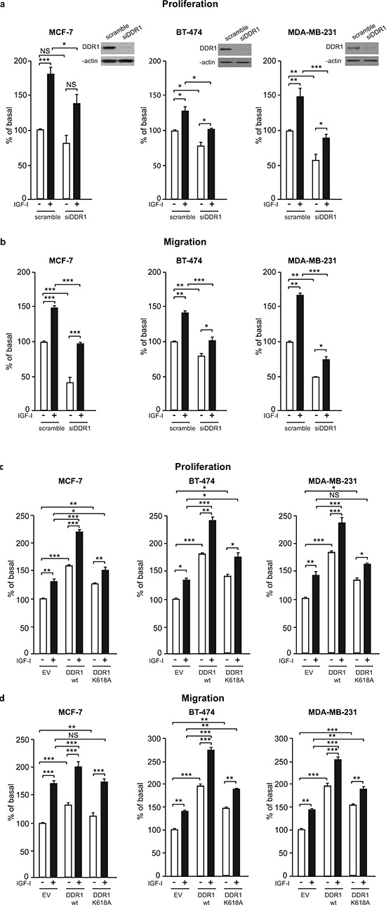
(a) Cell proliferation after DDR1 silencing. MCF-7, BT-474 and MDA-MB-231 breast cancer cells were transiently transfected with either a siRNA to DDR1 or scramble siRNAs. After 24 h, cells were grown in medium containing 2.5% of CS-FCS for 24 h and then incubated with or without 10 nM of IGF-I for further 48 h. Cell viability was evaluated by MTT assay. Values are expressed as percentages of untreated scramble oligo-transfected cells (basal) and represent the mean±SEM of three independent experiments in triplicate. NS, p > 0.05; *0.01 < p < 0.05; **0.001 < p < 0.01; ***p < 0.001; (untreated vs. IGF-I treated cells in scramble and siDDR1 conditions; untreated scramble vs. untreated siDDR1 cells; IGF-I treated scramble vs. IGF-I stimulated siDDR1 cells respectively). DDR1 silencing was confirmed for each cells lines by western blot analysis as shown on the right of each histogram. (b) Migration after DDR1 silencing. MCF-7, BT-474 and MDA-MB-231 breast cancer cells were transiently transfected as in (a) After 24 h, cells were grown in medium containing 0.1% of BSA for additional 24 h. Cells were then removed from plates with 0.01% trypsin and seeded on polycarbonate filters coated with 25 μg/mL fibronectin. Cells were allowed to migrate for 6 h (MCF-7 and MDA-MB-231) or 8 h (BT-474 cells) in response to 10 nM of IGF-I added to the lower chamber. Values are mean±SEM of three independent experiments done in duplicate and are expressed as percent of untreated scramble cells (basal). *0.01 < p < 0.05; **0.001 < p < 0.01; ***p < 0.001; (untreated vs. IGF-I treated cells in scramble and siDDR1 conditions; untreated scramble vs. untreated siDDR1 cells; scramble + IGF-I vs. siDDR1 + IGF-I). (c) Cell proliferation in DDR1-overexpressing cells. MCF-7, BT-474 and MDA-MB-231 breast cancer cells were transiently transfected with the wild type or kinase-inactive DDR1 mutant (DDR1/wt or DDR1/K618A) or the corresponding empty vector (EV). After 24 h, cells were grown in medium containing 2.5% of CS-FCS for 24 h and then incubated with or without 10 nM of IGF-I for further 48 h. Cell viability was assessed as in (A) Values are mean±SEM from three independent experiments in duplicate and are expressed as percent of untreated (EV) transfected cells (basal). *0.01 < p < 0.05; **0.001 < p < 0.01; ***p < 0.001; (untreated vs. IGF-I treated cells in EV, DDR1/wt and DDR1/K618A conditions; untreated EV transfected vs. untreated DDR1/wt or DDR1/K618A transfected cells; IGF-I treated EV transfected vs. IGF-I stimulated DDR1/wt or DDR1/K618A transfected cells). (d) Migration after DDR1 overexpression. MCF-7, BT-474 and MDA-MB-231 breast cancer cells were transiently transfected as in (c) Cell migration in response to 10 nM of IGF-I was evaluated as in (b) Values are mean±SEM of three independent experiments in duplicate and are expressed as percent of untreated (EV) transfected cells (basal). NS, p > 0.05; *0.01 < p < 0.05; **0.001 < p < 0.01; ***p < 0.001; (untreated vs. IGF-I treated cells in EV, DDR1/wt and DDR1/K618A conditions; untreated EV transfected vs. untreated DDR1/wt or DDR1/K618A transfected cells; IGF-I treated EV transfected vs. IGF-I stimulated DDR1/wt or DDR1/K618A transfected cells). (a–d) Statistical significance was calculated using one-way ANOVA followed by Bonferroni test.