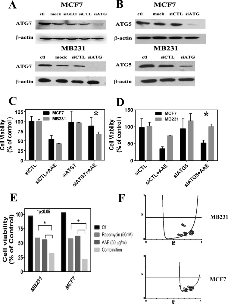
Figure 4. ATG knockdown blocks AAE induced autophagy and cell death.
A. & B. Immunoblotting of gene silencing by siRNA of ATG7 and ATG5 in MCF7 and MD231 cells. C. & D.: Percent of viable cells following various transfections with or without exposure to AAE. Transfected cells were harvested 48h after siRNA transfections, half the cell suspension used for protein estimation and immunoblotting. The other half of the harvested cells were incubated with 100 μg/ml AAE for 24h. Percent of live cells (viability) was determined after counting live and dead cells using trypan blue staining. Cell Viability was significantly rescued by autophagy inhibition of ATG7 and ATG5 compared to the background siRNA control group. E. Increased cytotoxicity in BrCa treated with both AAE and an autophagy inducer rapamycin. BrCa cells were treated either with AAE (25-75μg/ml) or rapamycin (10-50 nM) alone or with both drugs of various concentrations for another 72h. Cell viability was measured by MTT assay. Cell Viability decreased significantly in both treated MCF7 cells (*P < 0.05) and M231 cells (*P < 0.05) compared to AAE/rapamycin treated cells respectively. F. AAE and rapamycin synergistically induce cytotoxicity. The combination Index plot was generated using Compusyn (ComboSyn, Inc., Paramus, NJ, USA). Blue round circles indicate the data points in the drug combination treatment.

