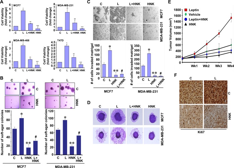
Figure 1. Honokiol diminishes the stimulatory effect of leptin on cell viability, anchorage-independent growth, invasion, migration and breast tumor growth in nude mice.
A. Breast cancer cells were treated with leptin and/or HNK as indicated and cell viability was examined by trypan blue dye exclusion assay. *p < 0.05 compared with untreated controls. Vehicle-treated cells are denoted with C. B. Soft-agar colony-formation of breast cancer cells treated with HNK and/or L as in A for three weeks. Histogram represents average number of colonies counted (in six micro-fields). *, P < 0.001, compared with Vehicle-treated cells (C); **, P < 0.005, compared with controls; #, P < 0.001, compared to leptin-treated cells. C. Analysis of Matrigel invasion of breast cancer cells treated as in A. Representative images are shown. The histogram shows mean of three independent experiments performed in triplicates. *, P < 0.005, compared with vehicle-treated controls (C); **, P < 0.001, compared with controls; #, P < 0.001, compared to leptin-treated cells. D. Spheroid migration assay of breast cancer cells in the presence of 100 ng/ml leptin (L), 5 μM HNK alone and in combination. The spheroids were photographed at 48h-post treatment. The results shown are representative of three independent experiments performed in triplicates. E. MDA-MB-231 cells derived tumors were developed in nude mice and treated with vehicle, Leptin, Honokiol (HNK) or Leptin + HNK. Tumor growth was monitored by measuring the tumor volume for 4 weeks. (n = 8-10); (P < 0.001). F. Tumors from vehicle (C), Leptin (L), HNK or HNK+L-treated mice were subjected to immunohistochemical (IHC) analysis using Ki-67 antibodies.

