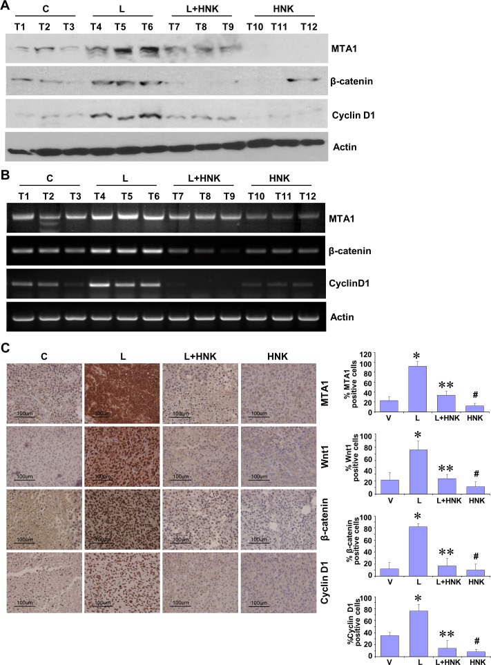
Figure 3. In vivo evidence for HNK-mediated inhibition of leptin-induced MTA1/Wnt1/β-catenin pathway in breast cancer cells.
MDA-MB-231 cells derived tumors were developed in nude mice and treated with Leptin (L), HNK, Leptin + HNK or vehicle (C). At the end of four weeks of treatment, tumors were collected for analysis. A. Total protein lysates from breast tumor samples were immunoblotted for MTA1, β-catenin, cyclin D1 expression levels. Actin was used as control. B. Total RNA was isolated from tumor samples and subjected to RT-PCR analysis. Expression of MTA1, β-catenin, cyclin D1 was analyzed. Actin was used as control. C. Breast tumors from each treatment group were subjected to immunohistochemical analysis using MTA1, Wnt1, β-catenin and cyclin D1 antibodies. Bar diagram shows quantitation of MTA1, Wnt1, β-catenin and cyclin D1 expression in tumors from each treatment group. Columns, mean (n = 5); *, P < 0.005, compared with control; **, P < 0.001, compared with leptin-treatment; #, P < 0.05, compared to untreated cells.

