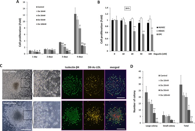
Figure 1. Dose-dependent suppression of EPC proliferation and colony formation by deguelin.

A. KSL cells (10.000 cells/well) were cultured in StemSpan media containing different concentrations of deguelin (n = 4/group). Relative cell numbers were measured by a CCK-8 assay at the indicated time points (*P < 0.05; **P < 0.001 vs. non-treated control). B. HUVECs, HDLECs were cultured in the EGM-2 media in different concentration of deguelin and relative cell numbers were measured by a CCK-8 assay at 24 h in compared to cultured EPCs as shown in A (n = 4). C. D. KSL cells were seeded in methylcellulose containing media containing deguelin at different concentrations (n = 4/group). Colony forming units were counted by blind investigators 12 days after seeding. Small- and large-CFUs were defined as focused clusters of rounded cells or as central core of round cells with elongated sprouting cells at their peripheries, respectively (left panel). Enlarged images of the boxed regions are shown in right. DiI-Ac-LDL was uptaken by these colonies and colonies were treated with FITC-conjugated anti-isolectin B4 antibody. C. Quantitative number of colonies were counted and graphed D. Three indepedent experiments were performed. *P < 0.05; **P < 0.01 vs. non-treated control. Scale bar = 500 μm.
