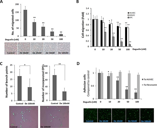
Figure 3. Effects of deguelin on EPC migration, adhesion, and tube formation.

A. A modified Boyden chamber assay was used with rhVEGF (20 ng/ml) as chemoattractant. EPC migration was identified as a red-purple color by staining with H&E after removing non-migrated cells (n = 4). B. The same migration assay experiment as in A using HUVEC, HDLEC, and cultured EPCs were performed in the presence or absence of deguelin (10–100 nM) in lower or upper chamber for 5 h (n = 3). C. GFP-expressing EPCs were co-cultured with HUVECs on Matrigel matrix for 8 h. Branching points and incorporated GFP-expressing EPCs were counted and graphed (n = 3). Representative pictures showing the incorporation of GFP-expressing EPCs into tube-like structures shown in below. D. EPCs were allowed to adhere to HUVEC monolayers for 3 h or to fibronectin for 30 min. The graph shows fold inductions of numbers of adherent cells versus non-treated control (n = 3). The lower panel shows representative pictures illustrating the adhesion of GFP-expressing EPCs to HUVEC monolayers. *P < 0.05; **P < 0.001 compared to control.
