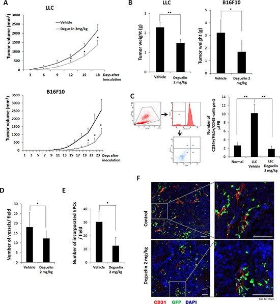
Figure 4. Effect of deguelin on tumor growth and vasculogenesis in vivo.

A. Mice were inoculated s.c. with LLCs or B16F10 cells and treated with deguelin (2 mg/kg body weight, n = 6) or vehicle (n = 6) every 2 days, and measured tumor volume. B. Tumor final weights of A. C. MNCs were isolated from peripheral blood by gradient centrifugation using Histopaque 1083 and stained with Alexa Fluor 647-conjugated anti-CD45, PE-conjugated anti-Flk-1 and FITC-conjugated anti-CD34 antibodies (n = 3). Cells were analyzed by flow cytometry. Debris and platelets were excluded by gating low FSC/SSC fractions. Circulating EPCs were defined as CD34+/Flk-1+/CD45− cells. D–F. Mice transplanted with BM-MNCs from GFP-expressing mice were s.c. inoculated with B16F10 cells and treated with deguelin as above (n = 6). Tumors were fixed and subjected to IHC. Staining for CD31 revealed tumor micro-vessel densities. D. Among the tumor microvessels, GFP-expressing EPC-derived vessels were counted. E. CD31 (red) and the differentiation of GFP-expressing EPCs into endothelial cells (green) were shown. F. Enlarged images of the boxed regions are shown in right. Each experiment was performed twice independently. *P < 0.05; **P < 0.005; ***P < 0.001. Scale bar = 100 μm.
