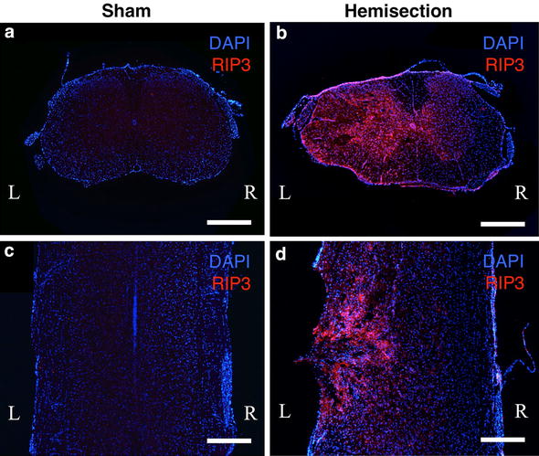
Fig. 1.

Immunohistochemical staining of RIP3 using spinal cord sections obtained 3 days after hemisection and the sham operation. The uninjured spinal cords in the sham operated mice (a, c) showed no obvious RIP3 expression. In contrast, a higher expression of RIP3 was observed on the injured side (L) than on the contralateral side (R) in the transverse (b) and coronal (d) sections after hemisection. Scale bars 500 μm
