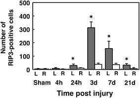
Fig. 3.

Number of RIP3-positive cells on the injured and contralateral sides at different time points. The number of RIP3-positive cells on the injured side (L) was significantly higher than that observed on the contralateral side (R) and in the sham group at 24 h and 3, 7 and 21 days. (*p < 0.05, n = 3 per each group). The values are presented as the mean ± SD
