
Figure 2. Connexin channels in cardiac ischemia.
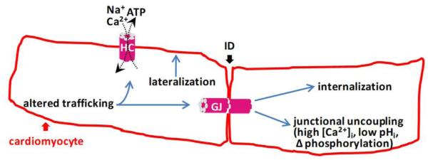
Connexins form gap junctions (GJ) that connect cardiomyocytes with each other at the site of cell-cell junction located intercalated disks (ID). They also form free unapposed hemichannels (HC) in the plasma membrane not incorporated in GJs. Ischemic conditions lead to junctional uncoupling and GJ closure as a result of [Ca2+]i elevation (Dekker et al., 1996; Peracchia, 2004; Xu et al., 2012), acidosis (Ek-Vitorin et al., 1996; Ek et al., 1994), altered phosphorylation status (Ek-Vitorin et al., 2006; Pahujaa et al., 2007) and other ischemia-related factors (Sanchez et al., 2011). In addition to this, connexins are remodeled as a result of processes that involve lateralization of connexin protein (Chkourko, et al., 2012; Kieken et al., 2009), altered trafficking (Remo et al., 2011; Smyth et al., 2010) and internalization (Duffy et al., 2004; Smyth et al., 2014; Sorgen et al., 2004). Most evidence comes from Cx43 which is a major connexin in ventricular cardiomyocytes that is also present in atria (in addition to Cx40). Cx43 hemichannels are normally closed but open in response to ischemia mimicking conditions (Kondo et al., 2000; Contreras et al., 2002), lowered redox status (Retamal et al., 2007; Saez et al., 2010), lowering of extracellular [Ca2+] (Li et al., 1996; Torres et al., 2012), moderate elevation (≤500 nM) of [Ca2+]i (De Vuyst et al., 2009; Ponsaerts et al., 2010; Wang et al., 2012a) and mechanical stress (Batra et al., 2014). They open with alkaninization, close with acidosis (Schalper et al., 2010) and close with above 500 nM [Ca2+]i elevation (Wang et al., 2012a). It is currently not clear how connexin remodeling impacts hemichannel function. GJ closure acts in a protective manner by limiting cell death spread to neighboring cardiomyocytes but may also lead to increased propensity for postischemic arrhythmogenesis because of heterogeneities in conduction velocity and consequent conduction delays. Hemichannel opening may lead to excessive entry of Na+ and Ca2+ and the loss of essential metabolites (ATP and others) from the cells (Saez et al., 2010).