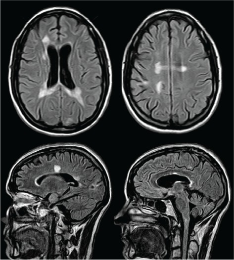
Figure 2.

Axial and sagittal fluid-attenuated inversion recovery images at the same levels as in Figure 1, 2 years later, showing resolution of the medullary lesion, coalescence of the periventricular lesions, and interval development of diffuse brain atrophy
