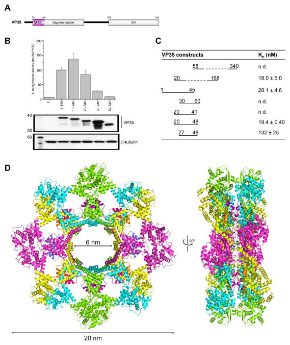
Figure 2. A peptide derived from EBOV polymerase co-factor VP35 (NPBP) binds ΔNPNTD with high affinity and specificity, related to Figure S1D, S2, S3, S4 and Table S1.
(A) Domain organization of EBOV VP35, including an oligomerization and IID domains. There is a highly conserved region N-terminal to the oligomerization domain, termed NPBP. (B) Impact of the VP35 N-terminus was evaluated by the Ebola minigenome assay. Ability of either wildtype or mutant VP35 proteins lacking various N-terminal sequences were tested and plotted as normalized minigenome activity, where wildtype VP35 activity was set to 100%. Below representative western blots show similar levels of VP35 protein expression. β-tubulin was used as loading control. (C) Summary of binding measurements by ITC between ΔNPNTD and VP35 N-terminal truncation mutants reveal that residues 20–48 are necessary and sufficient for high affinity binding. n.d., not determined. (D) The structure of the dual-octameric ring of ΔNPNTD in complex with VP35 NPBP peptide in a ribbon representation (left) and in a 90° rotated configuration (right). Each NPBP/ΔNPNTD complex in the asymmetric unit is colored yellow/orange (mol A/mol E), magenta/blue (mol B/mol F), green/purple (mol C/mol G), and cyan/pink (mol D/mol H).

