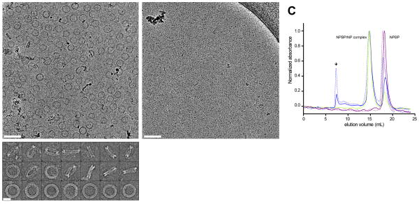
Figure 6. EBOV VP35 NPBP binding can dismantle oligomeric ring structures formed by NPNTD.
(A) Representative cryoEM images of NPNTD forming double ring structures, observed from top and side views. Scale bar: 100 nm (up), 20 nm (down). (B) Ring-like structures formed by NPNTD disappears upon addition of NPBP peptide just prior to cryoEM grid preparation. Scale bar: 100 nm. (C) Representative chromatograms of NPBP complex of NPNTD in 150 mM NaCl (blue dotted line) and 500 mM NaCl buffers (blue solid line), ΔNPNTD in 150 mM NaCl (green dotted line) and 500 mM NaCl (green solid line) from size exclusion chromatography. NPBP peptide alone is shown in 150 mM NaCl (purple dotted line) and 500 mM NaCl (purple solid line) buffer conditions. Location of the void volume for the Superdex 200 column is indicated with an arrow.

